Exercises after meniscus repair
For meniscus and other knee injuries, consult the best orthopedic surgeon in Jaipur, Dr. Jitesh Jain.
All about meniscus tears: Meniscus tear and treatment Anatomy:
The menisci are the wedge shaped cushion like discs (made primarily of type I collagen) that are sandwiched between the opposing thigh and leg bones. There are two menisci in the each knee joint, outer (lateral) and inner (medial). These menisci derive their nutrition primarily from synovial fluid (fluid that is present in the knee joint). Each meniscus has three parts namely a posterior horn, a body and an anterior horn. The inner meniscus is elliptical and larger in size than the outer meniscus which is more circular in shape and has the greater surface area.
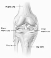
Why these menisci are important: Menisci are elastic structures and they act as shock absorbers in the knee joint and protect the joint cartilage (glistening surface of opposing thigh and leg bones). They are also important in keeping the thigh bone fit into leg bone. Menisci help in maintaining joint congruity and maximize contact area between leg bone (tibia) and thigh bone (femur).
What causes a meniscus tear?
In young patients meniscus tear are mostly due to twisting injury to the knee joint. In old persons these are mostly due to age related degenerative changes. Abnormalities of meniscus like abnormal shape (Discoid meniscus) or meniscal cyst also make menisci more prone to injury. Inner meniscus is less mobile than outer hence tear of inner meniscus are more common.
Can all meniscus tear be repaired?
Only peripheral 10-25% width of menisci has blood supply. This outer or peripheral zone is known as red-red zone. Inner 1/3rd width of menisci has no blood vessels. This is known as white-white zone. In between these two zones (red-white zone) a few blood vessels may present. Only tears of red-red zone, and a few tears of red-white have healing potential and can be repaired. A majority of meniscal tears are present in white-white zone and can’t be repaired. All tears of white-white zone and most of the tears of red-white zone need resection.
Signs and symptoms:
Pain and recurrent swelling are common features. Often a torn piece of meniscus entrapped in the joint gives a clicking sound and pain on knee bending as would occur in climbing up or down the stairs. At times, the torn part can be trapped in between the leg and thigh bones and may prevent leg bone from straightening. This condition is called locking of the knee. A locked knee may require manipulation by the patient or the doctor.
How meniscal tears are treated?
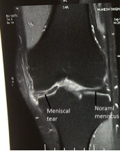
Meniscal tears are diagnosed by clinical examination and confirmed by MRI. Fresh tears, small tears and tears of peripheral red-red zone can be repaired. In meniscal repair torn parts are sutured together. Old tears, tears of white-white zone, degenerative tears and complex tears are usually trimmed away. All meniscal surgeries are key hole surgeries and done arthroscopically (minimally invasive).
Why these menisci are important: Menisci are elastic structures and they act as shock absorbers in the knee joint and protect the joint cartilage (glistening surface of opposing thigh and leg bones). They are also important in keeping the thigh bone fit into leg bone. Menisci help in maintaining joint congruity and maximize contact area between leg bone (tibia) and thigh bone (femur).
What causes a meniscus tear?
In young patients meniscus tear are mostly due to twisting injury to the knee joint. In old persons these are mostly due to age related degenerative changes. Abnormalities of meniscus like abnormal shape (Discoid meniscus) or meniscal cyst also make menisci more prone to injury. Inner meniscus is less mobile than outer hence tear of inner meniscus are more common.
Can all meniscus tear be repaired?
Only peripheral 10-25% width of menisci has blood supply. This outer or peripheral zone is known as red-red zone. Inner 1/3rd width of menisci has no blood vessels. This is known as white-white zone. In between these two zones (red-white zone) a few blood vessels may present. Only tears of red-red zone, and a few tears of red-white have healing potential and can be repaired. A majority of meniscal tears are present in white-white zone and can’t be repaired. All tears of white-white zone and most of the tears of red-white zone need resection.
Signs and symptoms:
Pain and recurrent swelling are common features. Often a torn piece of meniscus entrapped in the joint gives a clicking sound and pain on knee bending as would occur in climbing up or down the stairs. At times, the torn part can be trapped in between the leg and thigh bones and may prevent leg bone from straightening. This condition is called locking of the knee. A locked knee may require manipulation by the patient or the doctor.
How meniscal tears are treated?
Meniscal tears are diagnosed by clinical examination and confirmed by MRI. Fresh tears, small tears and tears of peripheral red-red zone can be repaired. In meniscal repair torn parts are sutured together. Old tears, tears of white-white zone, degenerative tears and complex tears are usually trimmed away. All meniscal surgeries are key hole surgeries and done arthroscopically (minimally invasive).
EXERCISE & REHAB
Rehabilitation after meniscus repair:
After meniscus repair you should not put weight on the operated limb for 4 weeks and your leg will be kept straight in a knee brace. You should be away from sports activities (jumping, cutting, twisting activities) for at least 6 months after surgery. Use walker or crutches for walk.
Week 1-2: you should apply ice every 2 hours for 20 minutes around operated knee for 1 week after surgery. This will help reducing edema and pain.
Exercises:
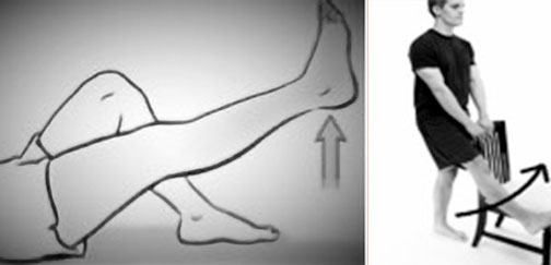
- Straight leg raise exercises (lying, standing): In lying down or standing position lift the leg (with brace) you want to exercise up to 45 degrees. Hold it there for 10 seconds and then repeat with other leg. Do 10 times with each leg and repeat two times a day.
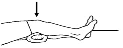
- Heel props: Place the heel on a rolled towel(without brace) and press into it for 5 seconds then allow the leg to relax into extension for 5 seconds. Do this exercise every waking hour for 10 minutes. This helps to achieve full extension (straight leg).

- Static quadriceps: In lying position keep a rolled towel under your knee and push into it(without brace). Hold for 10 seconds and then relax for 5 seconds. Do three sets of 10 repetitions

You can walk with walker support but don’t put weight on the operated leg (don’t touch operated leg with ground while walking in the first month).
Week 3-4
Continue all previous exercises.
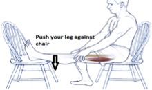
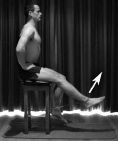
- Hamstring stretches: push your leg into chair as shown in the picture. Hold for the 10 seconds and repeat for 10 times. Do it twice a day.
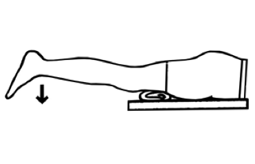
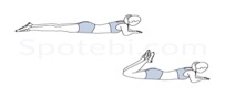
- Prone hangs to facilitate extension: lie down on the couch as shown in the picture, and keep the legs hanging at the edge of the table for 10 seconds. Repeat it for 10 times and do it twice a day.
- Straight leg raise in lateral position: In lying down position lift the leg (with brace) you want to exercise up to 45 degrees. Hold it there for 10 seconds and then repeat with other leg. Do 10 times with each leg and two times a day.
- Passive knee flexion (After 3 weeks): Remove brace and dangle your leg at the edge of the bed. Keep it there for a few seconds and then use other leg to straighten up operated leg. In such way allow pain free passive knee range of motion up to 90 degree.

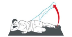
- Prone passive knee flexion (after 3 weeks): Use a Thera band (an elastic band) as shown in the picture and flex the knee in face down lying position. Bend your knee as much as you can. Hold for 30 seconds, then release. Repeat 3-5 times.


From weeks 3 onwards start passive knee flexion from 90 to 120 degrees. You can use Thera band/elastic band/Duptta to gain the flexion as shown in the picture below or when you hang the leg from the edge of the bed use push the operated leg with other leg to increase the range up to 120 degrees. Repeat 10 times and do it twice a day.

4-6 weeks: Now you wean off brace and start walking without brace. You can now put weight on the leg. You can continue using walker until you gain confidence to walk without support.
Continue all previous exercises:
- Heel raises/Toe raises: Hold for 5 seconds and slowly get down. Do it 3 times a day with 10 repetitions each time.

- Active knee extensions: Sitting in a chair, slowly straighten your knee from 90° to 30°, hold at 30° for several seconds, then bend back down to 90°. 3 sets of 10 repetitions.


- Hamstring curls: Lying on your front. Bend your knee up to 90° using your hamstring muscles. Slowly lower the foot to the floor again. 3 sets of 10 repetitions.
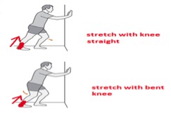
- Gastoc/soleus stretch: Lean towards a wall as shown in the picture and stretch your calf muscle. Don’t let your heel come off the ground. Hold the stretch for 15 seconds and repeat it for 10 times. Do it for 2 times a day. Repeat same exercise with bent knee.
- Stationary bicycle: you can start stationary bicycle in the 6th week after surgery. Start stationary bicycle 10 minutes a day. Gradually increase by 2 minutes per week till 20 minutes per week. Progress in this manner: high seat ½ circles forward/backward (pendulum exercises) to full circles – lower seat.

- Wall squats: Place feet at shoulder width. Take support of a wall and squat up to 30 degree. These are to be done at 3 angles of knee flexion. 30°, 60° and 90. Initially start with 30 degree and as you become comfortable increase the angle in the next few weeks. Hold for 10 seconds and then relax for 5 seconds. Do three sets of 10 repetitions.


Weeks 6-12
- Increase passive range of motion of the knee to full ( 0-135°)
- Gastroc/soleus stretch
- Hamstring stretch
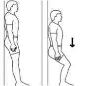
- Mini-squats: Squat up to 600. Don’t hold the flexed position and stand up smoothly. Do three sets of 10 repetitions.


- Hamstring curl (0-90°) against resistance: You can use elastic/thera band to do these exercises as shown in the picture below. Hold a chair or wall to support yourself. Thera bands come in different colours and their strength increase in this order: Yellow- red-green- blue- black –silver –gold.
So you start with yellow colour and gradually move on to gold. Repeat 10 times and do it twice a day.
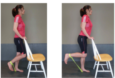
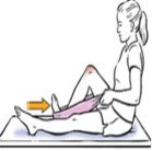
- Quadriceps strengthening exercises: This builds up your quadriceps (front muscles of thigh). Sit on a stool and pull thera band tied around ankle, as shown in the picture. Gradually increase strength of thera band. Do 10 minutes a day.
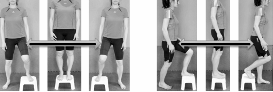
- Step-ups: After 8 weeks, you can do these exercises. These can be done forwards, backwards and from side to side. Start with a 10cm step and gradually increase its height to 30cm.
12-24 weeks: Continue doing all stretching and strengthening exercises. You should start balance training now.
– Single leg toe raises: Hold for 5-10 seconds and do two sets of 10 repetitions.
– Single leg balance, eyes closed: Hold for 5-10 seconds and do two sets of 10 repetitions.
– Single leg balance with various positions of head, arm and trunk: Hold for 5-10 seconds and do two sets of 10 repetitions.
24 Weeks on wards:
Agility Drills: Agility is one’s ability to change direction or position as quickly and effectively as possible. This is very important demand during sport activity. This gives confidence to get back to sports again and also help prevent re-injuries. Following are a few simple agility drills:
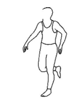
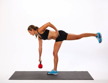
- Backward running: This is an effective drill particularly for sports requiring strength and endurance of the gluteal (buttock) and calf muscle (leg) groups. Start at slow speed and gradually increase the speed. Run for 50-60 meters and do 5-10 repetitions.
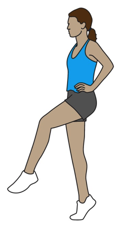
- High Knee Drills
While running lift knees as high as possible. Run for 30 seconds. Repeat two times a day.
- Vertical Jumping
Stand with your feet at a shoulder distance and bend your hip, knee and ankles until knee are flexed to 90 degrees. Now jump as high as you can. Land gently with knees slightly bent. Repeat 15 – 20 times.
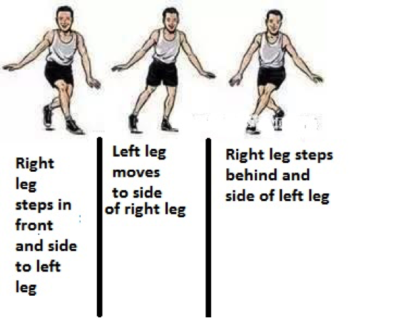
- Carioca
During carioca drill you move sideways at fast speed or run sideways. Move your right foot crossing front of your body and to side of left foot. Then move left foot to side of right foot. Now move right foot crossing behind the body and to side of left foot. Thus with every step you move sideways. Move/run to 30-40 meters.
Cutting Drills
- Run and Cut
Run at moderate speed for 10-15 meters straight, plant on the right leg and turn to change direction to left and then run again. Repeat 15-20 times.
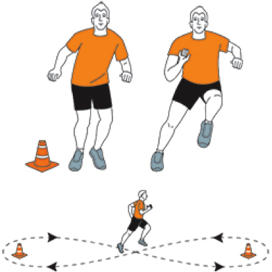
- Figure of Eight drill
Run a figure of eight pattern at moderate speed for 30-40 meters. Repeat it for 15 times. Gradually reduce the size of “8” and increase your speed as you progress. It is very helpful for developing “run and cut” skills in field sports.
Advanced weight training and sports specific drills are advised to maintain a
higher level of competition.
ABOUT THE AUTHOR

TESTIMONIAL

मेरा दाए पैर में चोट लगने के कारण टेढ़ा हो गया था जिसकी वजह से मुझे चलने में भी परेशानी हो रही थी. डॉ जितेश ने ऑपरेशन कर के इसे सीधा किया. में बिलकुल ठीक हूँ .

मेरी बेटी के घुटने में इन्फेक्शन होने के कारण वो दर्द की वजह से सो भी नहीं पाती थी. डॉ जितेश ने ऑपरेशन कर के इसे ठीक किया. बहुत धन्यवाद।

I remember that Dr. Jain came out from his clinic to see my mother because my mother was not able walk a single step. He did total knee replacement on both side and now my mother is walking without aid.
OPENING HOURS
| Monday – Friday | 17:00 – 20:00 |
| Sunday | OFF |